Predicting the risk of developmental dyslexia
(Patrycja Rusiak)
The problem of developmental dyslexia becomes a more and more common problem in Polish schools. Unfortunately, there is still a lack of standardised methods which would aid school teachers and psychologists in diagnosing this disorder in a clear and precise way. However, it should be realised that, in case of children with developmental dyslexia, the lack of proper and early therapeutic treatment may cause difficulties observed at all stages of school education and often it may be a serious obstacle in building proper relations with other people. Consequently, the lack of early diagnosis and, in the result, lack of proper action aiming at levelling possible difficulties may contribute to the child’s inferior start into their adult life.
Cerebellum Deficit
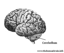
The concept of cerebellum deficit (Nicolson and Fawcett, 1994) assumes that the reason of all difficulties observed among dyslectic people lies in disorders in cerebellum construction and functioning. Cerebellum is responsible for language problems, balance difficulties, co-ordination as well as detection and learning of specific stimulants sequences. As the function of cerebellum is to automatisation skills acquired, therefore, deficiencies within this process cause difficulties in speech sounds articulation, which consequently causes problems on the level of phonological processing and later on problems with reading.
|
Apart from hearing difficulties, disorders within activities automatisation may cause problems in quick recognition of letters perceived, which may further aggravate the process of acquiring literary skills. Co-ordination and motor control disorders cause serious difficulties with writing. Cerebellum is often defined as the organ which supports so called inner speech. It is essential in reading education as it allows for matching phonemes with letters in a word read and further for connecting these speech sounds into a properly sounding word. The involvement of cerebellum in language processes is additionally evidenced by numerous connections of its right side with the temporoparietal areas of left cerebral hemisphere (Scott et al., 2001). Leiner et al. (1991) state that due to numerous connections with frontal cortex, where Broca’s speech area is located, cerebellum is involved in language skills acquisition and the performance of tasks connected with phonologic and semantic processing.
There is a series of evidence indicating the existence of numerous disorders within the cerebellum area among people with specific difficulties in reading and writing. Neuroanatomic examinations revealed anomalies in cells construction and their distribution within the dyslexics’ cerebellum area. (Finch et al., 2002). It also appeared that people with specific difficulties with writing and reading have untypical symmetry of both parts of this cerebellum and biochemical changes in this organ (Rae et al., 1998, 2002).They showed, that in case of good reader, during the tasks performance which required learning new reactions and the ones in which automatisation processes are involved, the cerebellum right side was strongly activated, whereas, in case of persons with dyslexia, frontal lobe areas responsible for conscious control during the performance of specific activities were activated, whereas the cerebellum was involved to a small extent.
There is a series of evidence indicating the existence of numerous disorders within the cerebellum area among people with specific difficulties in reading and writing. Neuroanatomic examinations revealed anomalies in cells construction and their distribution within the dyslexics’ cerebellum area. (Finch et al., 2002). It also appeared that people with specific difficulties with writing and reading have untypical symmetry of both parts of this cerebellum and biochemical changes in this organ (Rae et al., 1998, 2002).They showed, that in case of good reader, during the tasks performance which required learning new reactions and the ones in which automatisation processes are involved, the cerebellum right side was strongly activated, whereas, in case of persons with dyslexia, frontal lobe areas responsible for conscious control during the performance of specific activities were activated, whereas the cerebellum was involved to a small extent.
The aim of our study
The hypothesis of cerebellum deficit links developmental dyslexia with disorders in cerebellum construction and functioning. The aim of our investigation is to create tools facilitating early diagnosis of specific difficulties with reading and writing.
Tests aiming at defining possible cerebellum deficiencies include: a posturographic test, a sequential learning test and a mirror test and specialistic sight system test - optometric tests. On the basis of them it can be assumed if the children are in the group incurring the risk of appearing specific difficulties with reading and writing in their further education.
Tests aiming at defining possible cerebellum deficiencies include: a posturographic test, a sequential learning test and a mirror test and specialistic sight system test - optometric tests. On the basis of them it can be assumed if the children are in the group incurring the risk of appearing specific difficulties with reading and writing in their further education.
References:
- Finch, A J, R I Nicolson, and A J Fawcett. 2002. Evidence for a neuroanatomical difference within the olivocerebellar pathway of adults with dyslexia. Cortex 38:529-539.
- Nicolson, R.I. and Fawcett, A.J. (1994). Reaction times and dyslexia. Quarterly Journal of Experimental Psychology, 47A, 29-48.
- Rae, C., J. A. Harasty, T. E. Dzendrowskyj, J. B. Talcott, J. M. Simpson, A. M. Blamire, R. M. Dixon, M. A. Lee, C. H. Thompson, P. Styles, A. J. Richardson, and J. F. Stein. 2002. Cerebellar morphology in developmental dyslexia. Neuropsychologia 40 (8):1285-92.
- Rae, C., M. A. Lee, R. M. Dixon, A. M. Blamire, C. H. Thompson, P. Styles, J. Talcott, A. J. Richardson, and J.F. Stein. 1998. Metabolic abnormalities in developmental dyslexia detected by 1H magnetic resonance spectroscopy. Lancet 351 (9119):1849-52.
